
- Why us?
- Company
-
Services
Our Service
-
Technology
-
News & Resources
Clinic Resources
At CardioScan we see more than 600,000 heart studies annually! In review of 2020, we’ve gathered 50 of our most interesting cases and traces. Follow us to never miss a case.
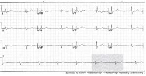
Some cardiologists are reluctant to identify something as complete heart block when there’s no AV dissociation, but that shouldn’t be the case. Dr Harry Mond explains why you shouldn’t be afraid to call something a complete heart block when it appears that way, what clues. Read more.

Cardiac technicians and those that report regularly on Holters may be familiar with cases like this, but the irregularities revealed in this patient are rarely reported on. Dr Harry Mond once again shares insights into another interesting trace, and what’s really going on. Read more.
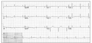
Working out whether an ECG is showing sinus rhythm with underlying artefact or atrial fibrillation is something that comes up regularly, and is the most common mistake made in ECG reporting. Dr Harry Mond explains how by digging a little deeper, differentiating between sinus rhythm. Read more.

The surprising trace that made even the most experienced cardiologist take pause. In this case of a ‘rebel without a pause’, Medical Director Dr Harry Mond examines Wenckeback sequences and the confusion with complete heart block that can sometimes occur. Read more.
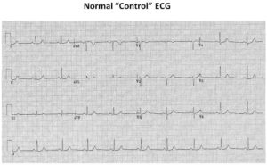
This week, Assoc Prof Harry Mond looks at the upside down ECG, and the puzzling trace that resulted. Each week, as our Director of Medicine he shares the real findings of unusual traces discovered at CardioScan, and what they reveal. Read more.

A recent Holter monitor report caused spirited debate amongst the team about whether it showed an atrial ectopic conducted with aberration, or a ventricular ectopic. CardioScan’s Medical Director, Assoc Prof Harry Mond, analyses the ECG and explains who came to the correct conclusion, as well. Read more.

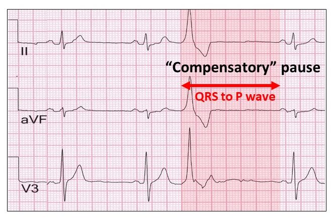
Compensatory pauses often follow atrial ectopics, but they can cause some confusion. In his latest case study, our Medical Director Dr Harry Mond clarifies the different types of compensatory pauses that can occur, as well as the typical characteristics of each compensatory pause as they. Read more.

A recent ECG was reported as sinus rhythm with intermittent bundle branch block – but this diagnosis was incorrect. CardioScan’s Medical Director Dr Harry Mond discusses the identifying factors in the ECG, and how he reached his diagnosis of an idioventricular rhythm with isorhythmic AV. Read more.

As a rare find Wandering Atrial Pacemaker can be mistaken for marked sinus arrhythmia with unifocal atrial ectopics. Here, we look at the tell-tale characteristics that set them apart in another interesting case study by Medical Director Dr Harry Mond. Read more.

All pacemaker companies have programmable algorithms to prevent ventricular pacing, but this can cause issues with Holter monitor test results. Our Medical Director, Dr Harry Mond, explains how pacemakers can confuse Holter monitor tracings, and how to correctly identify these algorithms on ECG patterns. Read more.

Reversed Wenckebach occurs when there is sequential shortening of the PR interval, and can require a permanent pacemaker in certain instances. In this latest case study, we take a look at examples of reversed Wenckebach, and how to recognise the rare ECG finding. Read more.

Not all ECG recordings are straightforward, as illustrated by this “bizarre” ECG. In this latest edition in our clinical case studies series, our Medical Director Dr Harry Mond explains how he assessed an ECG he was asked to look at, and how eliminated incorrect solutions. Read more.

Overnight Wenckebach AV block is a common finding in young people and is usually found in the presence of sinus bradycardia/sinus slowing. In this latest edition in our clinical case studies series, we look at how to identify atypical Wenckebach AV block, and how it’s. Read more.
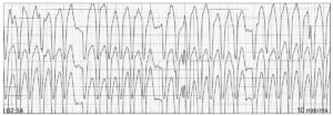

The presentation of pseudo Wolff-Parkinson-White Syndrome recently had an international customer return a report with a note pointing out that we had missed the diagnosis of intermittent pre-excitation – the Wolff-Parkinson-White Syndrome. But as Assoc Prof Harry Mond explains in this latest cardiac case study. Read more.

Reported as dual chamber pacing, this case study needed closer examination. With obvious atrial pacing, the question of ventricular pacing remained. Assoc Prof Harry Mond details the characteristics that reveal the correct diagnosis, and why this should not be confused with pacemaker malfunction. Read more.

This ECG was reported as complete heart block, and at first glance it sure looks like it. But a closer examination revealed the ventricular response was irregular. In this latest case study Assoc Prof Harry Mond explains two P wave patterns that reveal the rhythm. Read more.
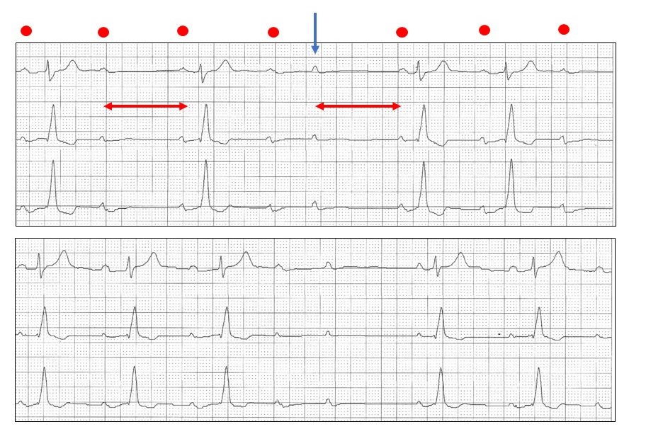

Sinus arrhythmia is unusual in a 63-yr-old but we were asked to amend a ‘normal’ result to reflect this diagnosis. With classical examples of the pattern in under 30s and showing how NOT to confuse it with atrial ectopy, Assoc Prod Harry Mond shows how. Read More.

Handed this ECG, our Medical Director Assoc Prof Harry Mond was asked if it was an example of a rate adaptive pacing, which uses changes in transthoracic impedance to increase the pacemaker rate in response to physiologic demand. Read more.

Seen for the first time, the pattern in this 24hr histogram gives instantly recognisable clues about what is happening, even without looking at the tracings. Read more.
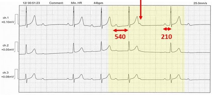
The Holter monitor tracings of a 44-year old male caused a lot of excitement at CardioScan this week. With 12 bradycardia episodes overnight, it was initially considered Wenckebach AV block. While clinically, rather than visually, correct, it nevertheless, did not fulfil the footprints of Wenckebach. Read more.
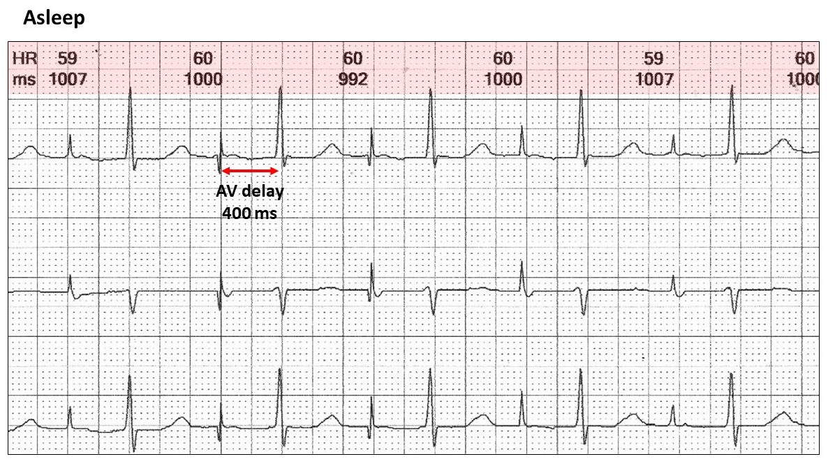
First thoughts on this Holter tracing was artefact, but the native rhythm showed a regular pattern of irregularity! Taking a close look at overnight tracings when the rhythm would be slow and the “artefact” less likely, revealed what was really going on. Read more.

In this rare case, we see two very unusual and critical factors that together lead to atrial oversensing and “apparent” violation of the lower rate limit; a very narrow zone of open atrial sensing and far-field R wave sensing. Read more.

In this first of his Fun With ECG series, our MD Assoc Prof Harry Mond looks at this bizarre atrioventricular (AV) delay. Look closely at the ECG. The AV delay timings don’t make sense. Read more.

With a pause about twice the cycle length of the R to R intervals and no P, QRS or T waves, this case tackles one of the most difficult explanations in ECG interpretation and demands a revisit to the fundamentals or “footprints” of Wenckebach AV. Read more.
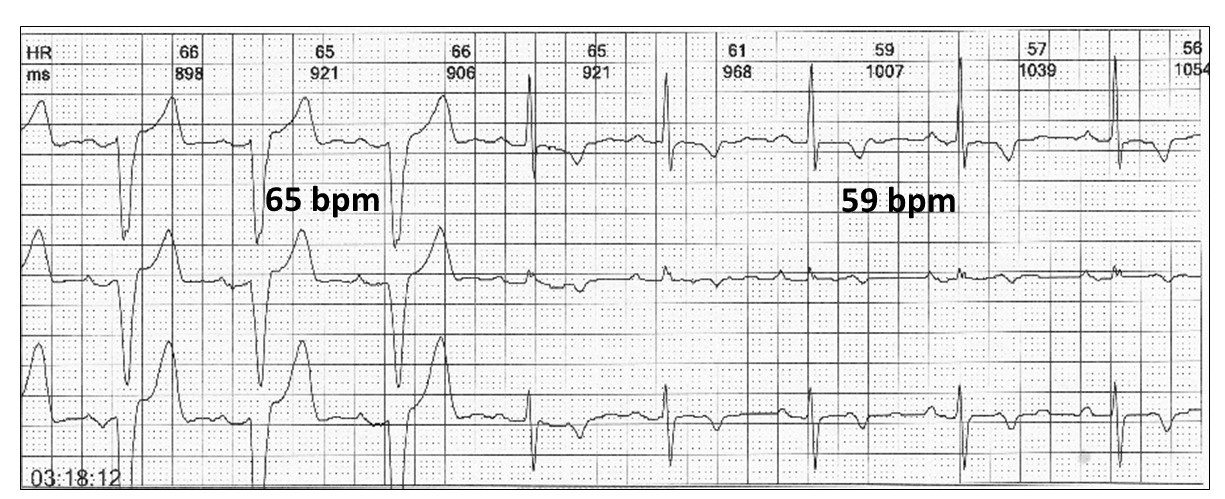
Exploring another ventricular aberration this week, we look at rate dependent bundle branch block, its defining characteristics, a number of important differential diagnoses and how to avoid serious misdiagnosis. Read more.

This week MD Assoc Prof H Mond examines the similarities between the characteristics of artefact vs non-atrioventricular Wenckebach blocks and the importance of recognising footprints for correct diagnosis. Read more.

A ‘bizarre’ case of dual chamber pacing with an 80ms AV delay, called us for a second opinion! With examples of a) Atrial pacing at 50 bpm and prolonged AV delay b) Wenckebach AV block; & Ventricular paced beat after 2 secs, we deep dive. Read more.
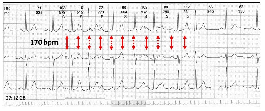
Assoc Prof Harry Mond identifies a plethora of insights that come with studying Wenckebach sequences. This week, a definitely not boring look into atrioventricular (AV) and non-AV types, and how it’s all in the timing! Read more.

Assoc Prof Mond shares this case to delve into unusual and classical patterns of twisted leads in 12-lead ECG, including the double twist, and shares the tell-tale footprints of all. Read more.
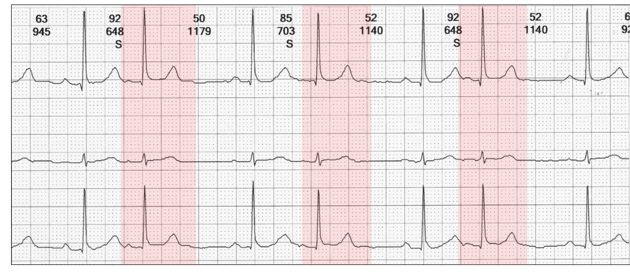
In part one of a two part series, Assoc Prof H Mond explores exotic atrial ectopy in all its forms, including the anatomy and characteristics of 12 different examples. Read more.

In the final part of this 2-part series, Assoc Prof H Mond further explores the different forms of atrial ectopy including the atrial couplet, triplet, run & non-conducted types. Read more.
34. Unusual ventricular ectopy

A controversial one this week: AFib with ventricular bigeminy. Or is it? Exploring trigeminy, ventricular parasystole, pseudo-ventricular parasystole and other possibilities, this one is sure to divide opinion. Read more.

A look at exotic ventricular ectopy this week, with 8 different examples, their footprints and the veritable kaleidoscope of findings that may result. Read more.

In the final part of this 2-part series, we explore even more exotic ectopy, including ventricular couplets, interpolation, unipolar bigemilets, trigemilets and more. Read more.
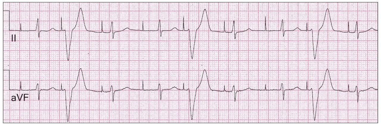
Often asked to review cardiac pacing ECGs, this one was presented as intermittent failure of ventricular capture. Revisiting atrial pacing and the tell-tale clues, we’re able to make the correct diagnosis. Read more.
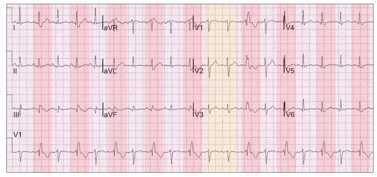
This week Assoc Prof Mond discusses the not so boring bundle branch block while exploring some puzzling occurrences including alternating bundle branch block and bidirectional rhythm. The clue is in the rhythm strip. Read more.
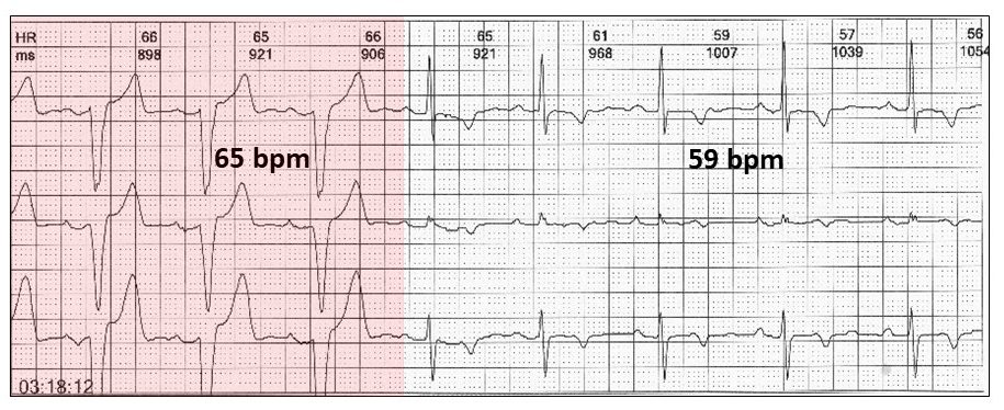
Last week we proved not all bundle branch blocks are boring. Now we delve even deeper into mistaken rhythms, common misdiagnosis and those rarely talked about, including the crochetage sign. Read more.
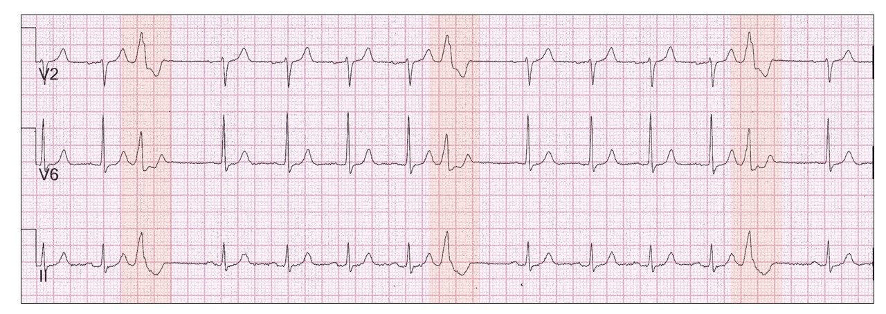
Something rarely seen in Holter monitoring, the ventricular pentageminy with ventricular ectopics every 5th beat, and other curious rhythms! Read more.
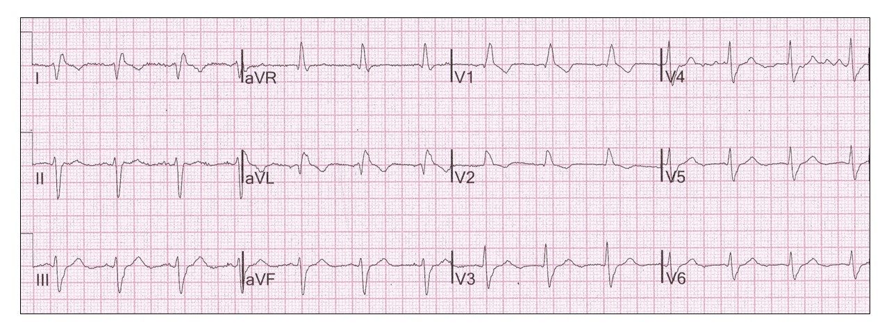
This week, Assoc Prof Mond discusses abnormalities commonly missed by physicians when reporting on ECGs, with a focus on reversed arm leads and how to avoid serious misdiagnosis. Read more.
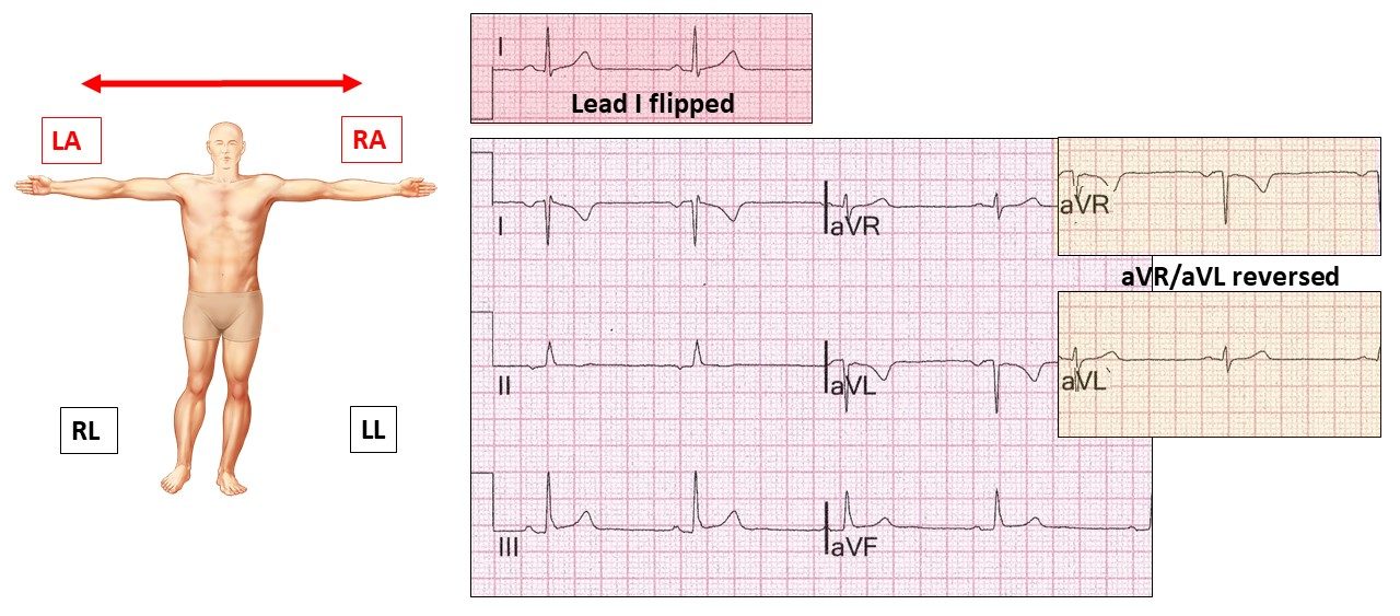
Last week some of you suggested dextrocardia in response to our case of reversed arm leads. This week shows that with dextrocardia the chest leads are very different and how to avoid misdiagnosis. Read more.

Can ECGs cause headaches? Well this one might! Leading on from our last ECG topic, this week we further explore twisted leads but involving all limbs with “leg leads on arms” & “arm leads on legs” & the major defining footprints. Read more.
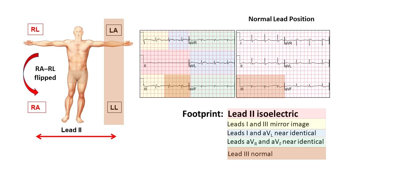
After recently discussing the two most common variations of reversed arm and leg leads, we now explore combinations of Right Arm-Right Leg Reversed, including the double twists, & all the tell-tale footprints. Read more.



In 49+ years as a practicing cardiologist, Assoc Prof Harry Mond has published 260+ published manuscripts & books. A co-founder of CardioScan, he remains Medical Director and oversees 500K+ heart studies each year.
Download his full profile here.